Researchers in the United States have developed artificial intelligence tools that may help physicians assess how seriously ill a cancer patient is by analysing a single photograph of their face. According to BBC News, the system was trained on millions of images of healthy faces alongside 35,000 photographs of cancer patients, enabling it to estimate survival prospects by examining specific facial features, particularly around the eyes and nose.
While the technology remains experimental and unsuitable for immediate hospital deployment, the research team emphasises that their objective is to support doctors in selecting appropriate treatments rather than replacing clinical judgment. The findings appear in the Lancet Digital Health journal.
The development builds on earlier progress in AI-assisted medical diagnosis. A Harvard study revealed AI outperforms doctors in emergency triage diagnoses and treatment planning, demonstrating the growing capability of machine learning in clinical settings, though human oversight remains essential. Similarly, the world's first patient underwent brain surgery with real-time AI assistance at University College London Hospital, showing how these tools can augment rather than replace physician expertise.
Which facial features matter most for survival prediction?
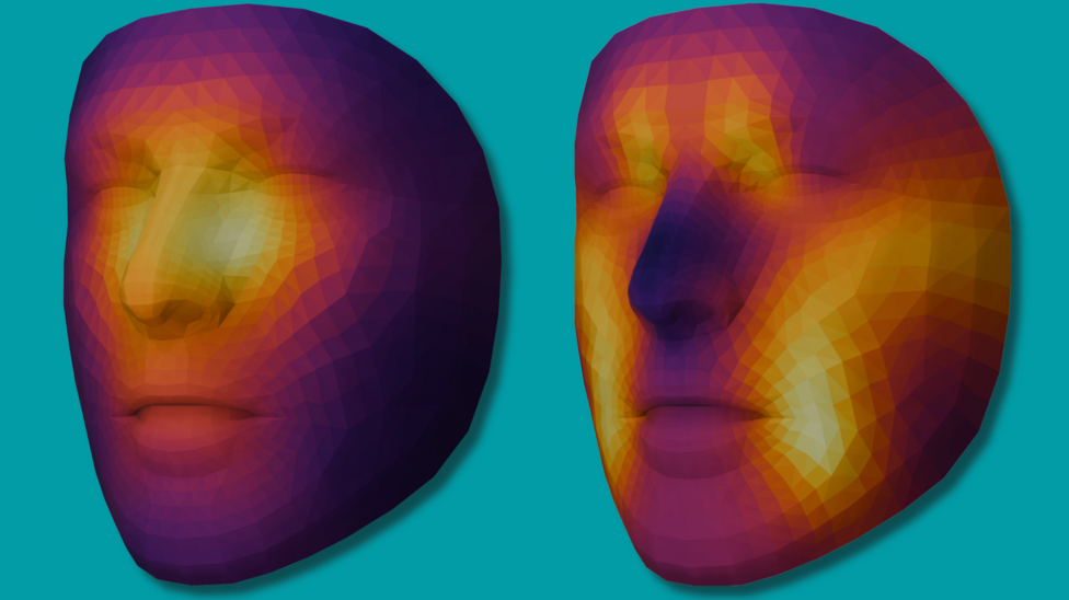
The 'FaceSurvival' AI tool identifies the area beneath the eyes and around the bridge of the nose as the most critical facial regions for estimating cancer survival outcomes. These metrics proved applicable across numerous cancer types of varying severity, including cancers that had metastasised to the bones or brain.
The research cohort comprised cancer patients primarily from the United States and the Netherlands, with ages ranging from children to those in their 90s, though most participants were in their 50s or 60s. According to SciMex, a newer model called FAHR-Face was trained on more than 40 million facial images and demonstrated striking predictive power: among patients assigned the highest risk scores, 64 of 122 died within 90 days, compared with only 20 of 975 patients assigned the lowest risk scores.
The same research found that cancer patients who appeared at least five years older than their chronological age faced a 21% higher risk of death during an average four-year follow-up period, while those appearing at least five years younger had a 17% lower risk. This suggests that apparent facial aging may serve as a marker of underlying health status and biological reserve.
How does the biological age tool work?
Alongside the survival prediction system, researchers are developing the 'FaceAge' tool to estimate biological age—how old someone appears visually—as distinct from their actual chronological age. The system identifies skin characteristics below the nose, including nasolabial folds and marionette lines, along with forehead creases, crow's feet around the temples, and cheek texture as reliable indicators of aging speed.
Dr Raymond Mak, the lead researcher from Mass General Brigham in Boston, noted that the tool demonstrated
"surprisingly consistent" results across common real-world photographs despite variations in pose, lighting, facial expression and make-up, and remained
stable across most tested plastic surgery procedures, with only a small shift observed after rhinoplasty.
Certain lifestyle factors accelerate the aging process visible in facial features, including heavy alcohol consumption, drug use, and smoking. A follow-up analysis of 2,276 cancer patients found that faster facial aging between successive clinical photographs was associated with worse overall survival, suggesting the tool might track health changes over time.
Why do doctors need this tool?
Physicians currently examine patients and conduct comprehensive assessments to determine illness severity and prognosis. Dr Mak explained the motivation behind the research:
As the population ages, physicians need better ways to estimate physical reserve and frailty before recommending intensive treatments such as radiation, surgery or systemic therapy. Our goal is to make face photograph-based health assessment more quantitative, consistent and scalable so it can support personalised treatment decisions rather than replace physician judgment.
The AI tool, which has a patent application pending, is explicitly designed to assist doctors in their existing work rather than substitute for their expertise and clinical experience.
What limitations remain?
Researchers acknowledge significant gaps in their current work. They stress the need for substantially more data and facial photographs, particularly from diverse ethnic groups, to establish how reliably the tools function across different populations. The systems remain under active development.
Prof Miroslaw Bober, an expert in video processing and machine learning at the University of Surrey in the UK, cautioned that any assertions about the tool's capacity to monitor aging or lifestyle changes over time would be
premature. He characterised the work as
a promising proof of concept, but it is not ready for clinical use, and noted that
the tool still needs testing against doctors' own judgement and across different hospitals and patient groups.
Bober also highlighted a fundamental distinction:
Predicting an outcome is not the same as explaining it.This distinction matters because understanding why a prediction is made—the biological mechanisms—differs from simply forecasting a result.
According to Becker's Hospital Review, the two newer models' predictions were only weakly correlated with each other, though combining them improved the survival model's fit in external validation cohorts, suggesting complementary rather than redundant information.
What happens next?
The research team is inviting members of the public to participate in the study and receive a personalised biological age estimate by uploading their own facial photograph. Mass General Brigham has announced that clinical trials are being launched to evaluate how well FaceAge-related measures predict outcomes in real-world settings. Researchers emphasise that further investigation is necessary before these newer models can support treatment decisions in clinical practice.